
Connect alveolar structure with diffusion between air and blood. The images provide a basis for explaining the exchange surface and the gradients maintained by ventilation and blood flow.
Choose from five images below. Open an image for a closer look, download its original PNG, or download all five as a ZIP.
Use these visuals in your lesson
Trace oxygen and carbon dioxide in opposite directions. Link each structural feature to a consequence for diffusion instead of listing adaptations without explanation.
A question to discuss
How do ventilation and blood flow help maintain diffusion gradients?
Alveolar gas exchange: five teaching visuals

Gas exchange
Oxygen moves from alveolar air into blood while carbon dioxide moves in the opposite direction.
Download original PNG
The exchange barrier
A close-up identifies alveolar epithelium, fused basal lamina and capillary endothelium.
Download original PNG
Partial-pressure gradients
Typical resting sea-level values illustrate the gradients for oxygen and carbon dioxide exchange.
Download original PNG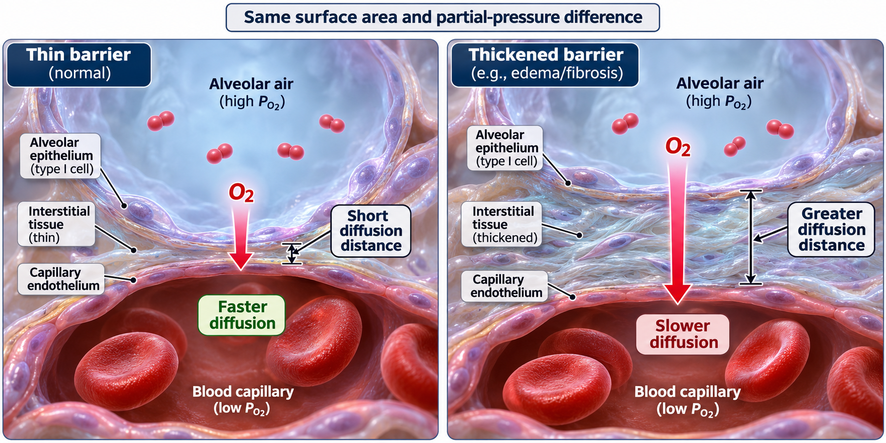
Barrier thickness
With surface area and pressure difference held constant, a thicker barrier slows diffusion.
Download original PNG
Alveolar tissue
A tissue cutaway and enlarged inset connect alveolar air spaces with the thin blood–air barrier.
Download original PNG