
Help students move from recognising an animal cell to explaining how its structures work together. These five views offer different amounts of visual detail, so you can choose the image that fits your explanation.
Choose from five images below. Open an image for a closer look, download its original PNG, or download all five as a ZIP.
Use these visuals in your lesson
Begin with the labelled overview, then use an unlabelled view for retrieval. Ask students to connect each structure to a function rather than simply naming it.
A question to discuss
How would a cell that secretes proteins make use of its nucleus, ribosomes and membranes?
Animal cell: five teaching visuals
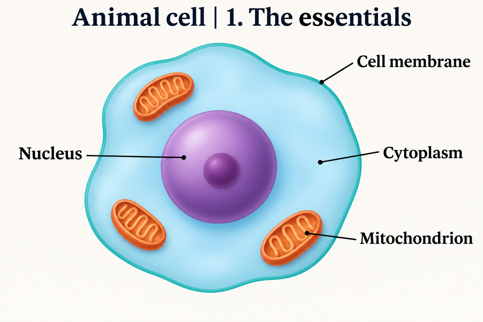
The essentials
A simple, labelled overview for introducing the main cell structures.
Download original PNG
Inside the cell
A labelled view of the organelles and the spaces around them.
Download original PNG
Cellular architecture
A richer cutaway that brings the internal structures into focus.
Download original PNG
Membranes and organelles
A detailed view for looking more closely at the cell’s organisation.
Download original PNG
The cellular world
An intricate illustration with layers of internal cellular detail.
Download original PNG