
Build an explanation of membrane structure from the phospholipid bilayer to proteins, surface carbohydrates and transport routes. These images connect the fluid mosaic model to what membranes do.
Choose from five images below. Open an image for a closer look, download its original PNG, or download all five as a ZIP.
Use these visuals in your lesson
Have students trace the route of a small nonpolar molecule and then an ion. Ask them to explain why the two routes differ rather than memorising a diagram.
A question to discuss
Why can oxygen cross the lipid bilayer more readily than a sodium ion?
Cell membrane: five teaching visuals
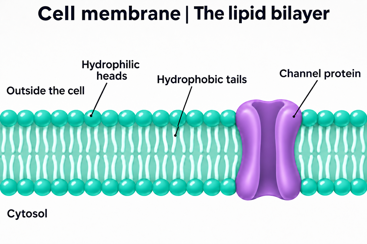
The lipid bilayer
Introduce the arrangement of phospholipids in a cell membrane.
Download original PNG
The fluid mosaic model
Explore the proteins and lipids shown in a fluid mosaic membrane model.
Download original PNG
A molecular landscape
A detailed membrane view for discussing its molecular organisation.
Download original PNG
Two different faces
Compare the extracellular surface with the cytosolic side of this membrane.
Download original PNG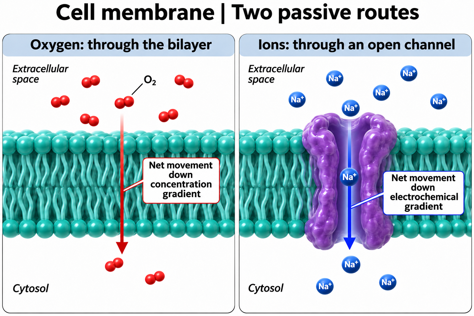
Two passive routes
Compare oxygen diffusion through the bilayer with ion movement through an open channel.
Download original PNG