
Follow semiconservative replication from strand separation to complementary synthesis and fragment joining. The collection makes strand direction and the differences between leading and lagging synthesis explicit.
Choose from five images below. Open an image for a closer look, download its original PNG, or download all five as a ZIP.
Use these visuals in your lesson
Have students mark 5′ and 3′ ends before tracing synthesis. Use the fragment-joining view to distinguish primer replacement from the final sealing of a nick.
A question to discuss
Why is one new strand assembled in fragments at a replication fork?
DNA replication: five teaching visuals

One old strand in each copy
Semiconservative replication leaves one parental strand in each new DNA molecule.
Download original PNG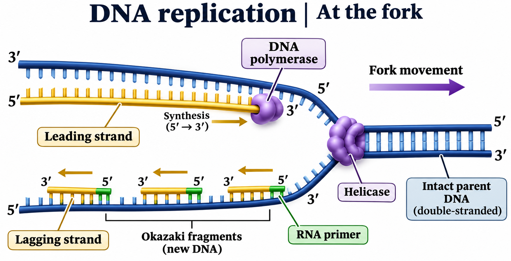
At the fork
Leading and lagging strands, helicase, primers and 5′ to 3′ DNA synthesis.
Download original PNG
Adding one nucleotide
A complementary nucleotide joins the growing strand, with pyrophosphate released.
Download original PNG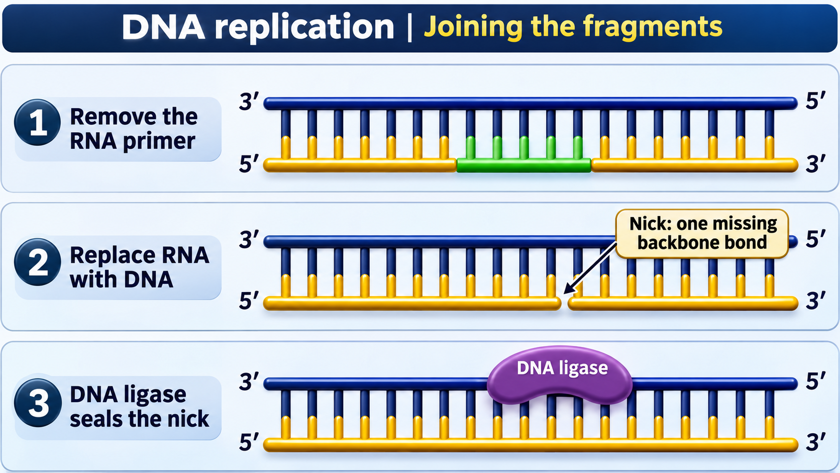
Joining the fragments
Primer removal, DNA replacement and ligase sealing a remaining nick.
Download original PNG
Opening the double helix
An illustrative view of helicase and single-strand binding proteins at a replication fork.
Download original PNG