
Use molecular representations derived from the deposited 1BNA DNA structure to examine DNA at different levels of detail. The images move from a sugar trace to atoms, space-filling views and a base-pair close-up.
Choose from five images below. Open an image for a closer look, download its original PNG, or download all five as a ZIP.
Use these visuals in your lesson
Compare what each representation reveals and what it leaves out. Students can use the chain-coloured view to distinguish the two strands before looking at individual atoms.
A question to discuss
What is easier to see in a space-filling model than in an atom-and-bond model?
DNA: five teaching visuals
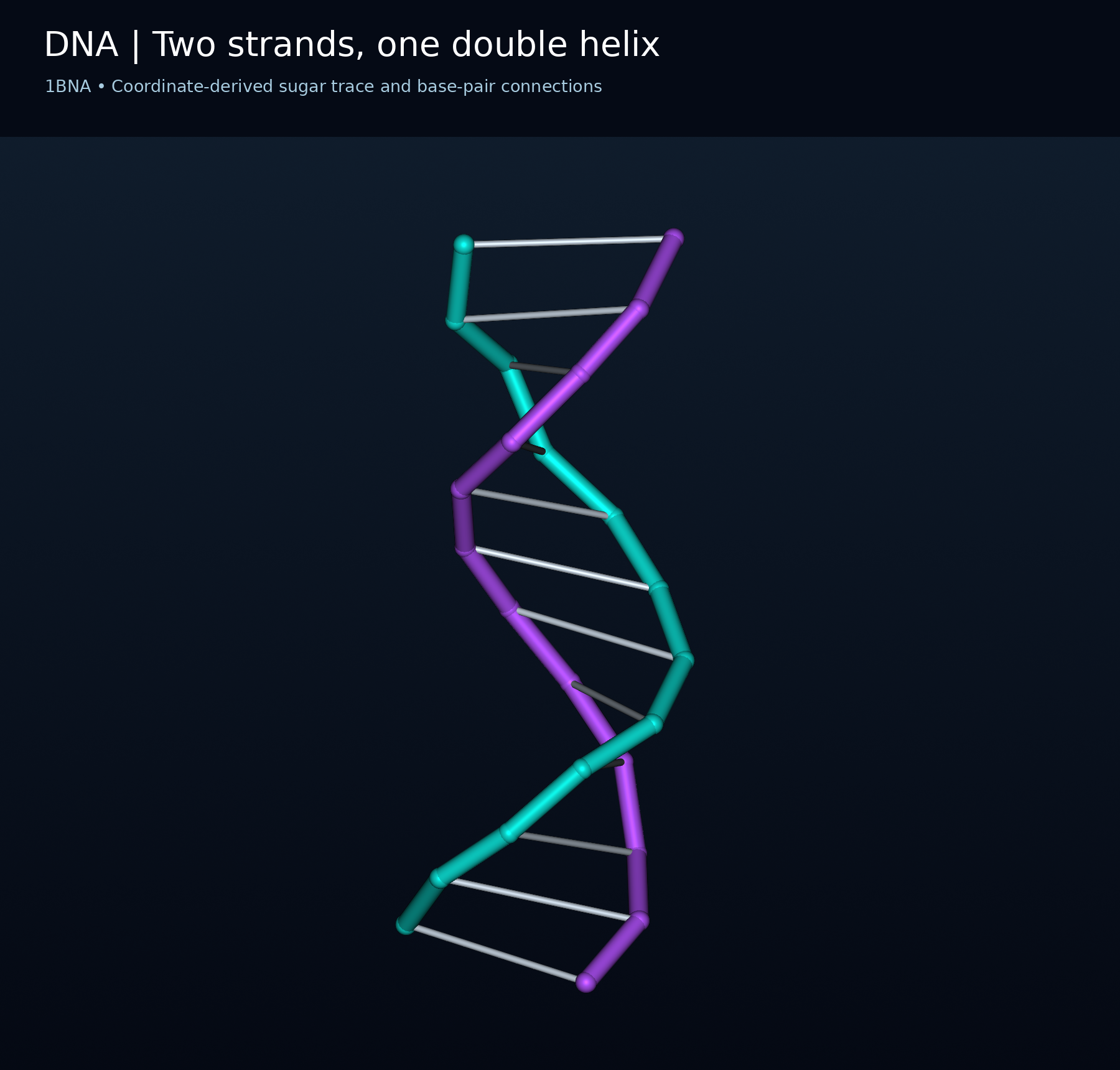
Two strands, one double helix
Coordinate-derived sugar trace and base-pair connections from DNA structure 1BNA.
Download original PNG
The atomic framework
Deposited non-hydrogen atoms of 1BNA, with the two DNA chains coloured separately.
Download original PNG
Atoms and covalent connections
A 1BNA atom-and-bond view with carbon, nitrogen, oxygen and phosphorus distinguished by colour.
Download original PNG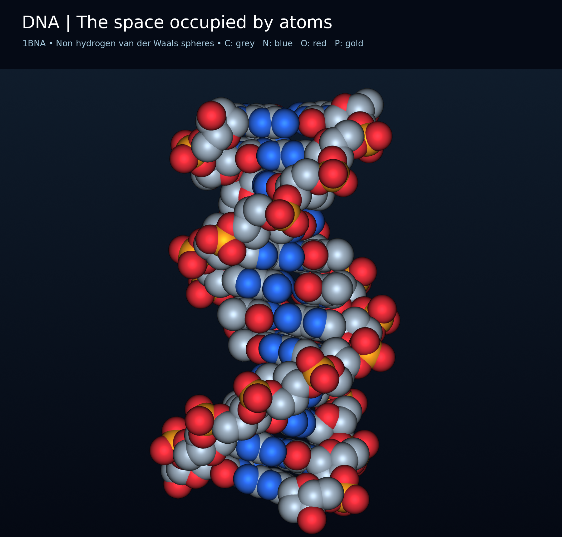
The space occupied by atoms
Non-hydrogen van der Waals spheres rendered from 1BNA coordinates.
Download original PNG
An adenine-thymine pair
Base atoms of 1BNA residues A6 and T19; hydrogen atoms and hydrogen-bond lines are omitted.
Download original PNG