
Explore the uptake of material through membrane invagination and vesicle formation. The collection supports comparison of routes and the connection between membranes and intracellular transport.
Choose from five images below. Open an image for a closer look, download its original PNG, or download all five as a ZIP.
Use these visuals in your lesson
Trace the membrane as well as the material being taken in. Ask students to explain how uptake into a vesicle differs from movement through a membrane protein.
A question to discuss
Why is material inside an endocytic vesicle not simply free in the cytosol?
Endocytosis: five teaching visuals

Membrane to vesicle
Three stages show the plasma membrane enclosing extracellular cargo in an internal vesicle.
Download original PNG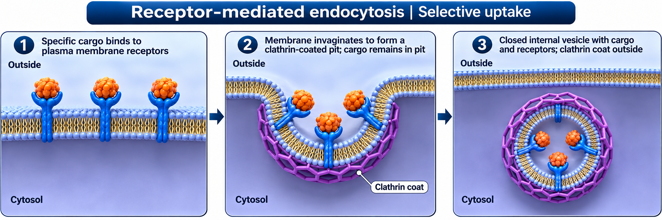
Receptor-mediated uptake
Receptors select cargo as a clathrin-coated pit forms and buds into the cell.
Download original PNG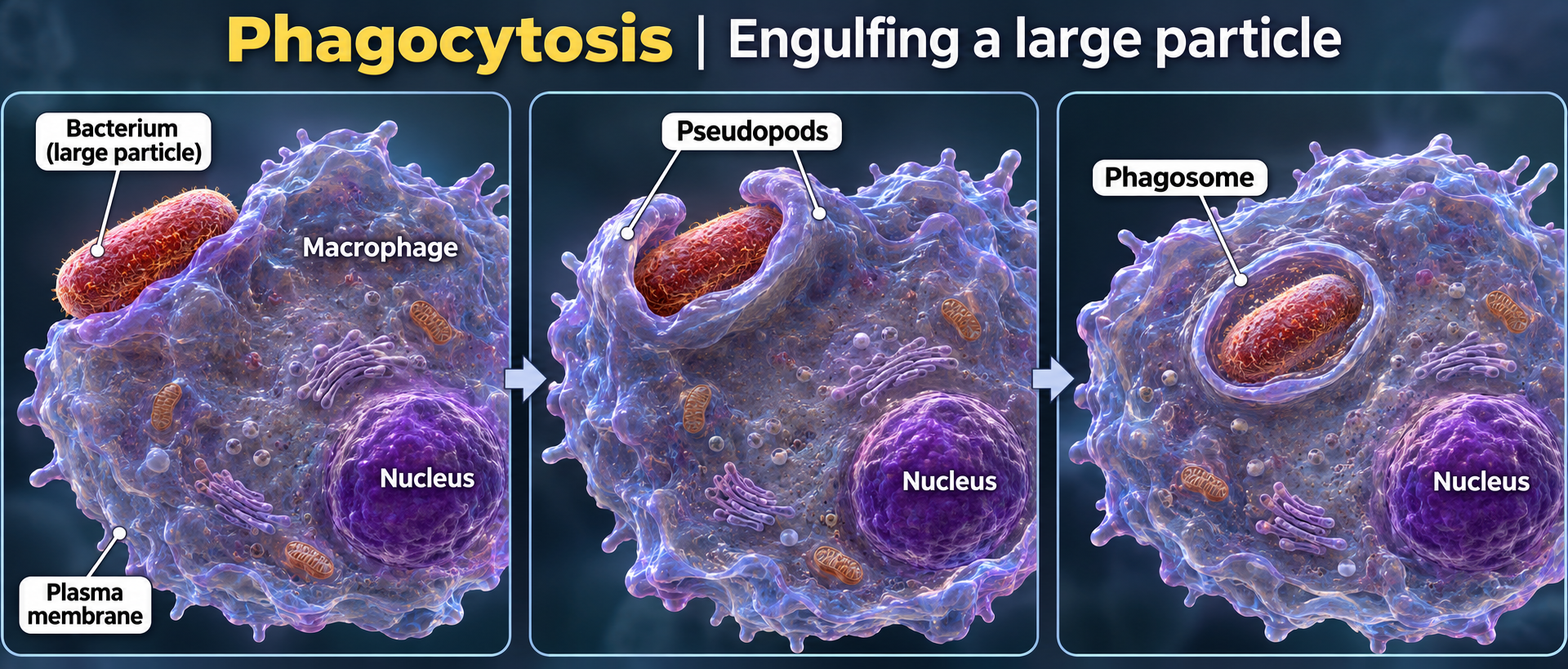
Phagocytosis
A macrophage extends pseudopods around a bacterium and encloses it in a phagosome.
Download original PNG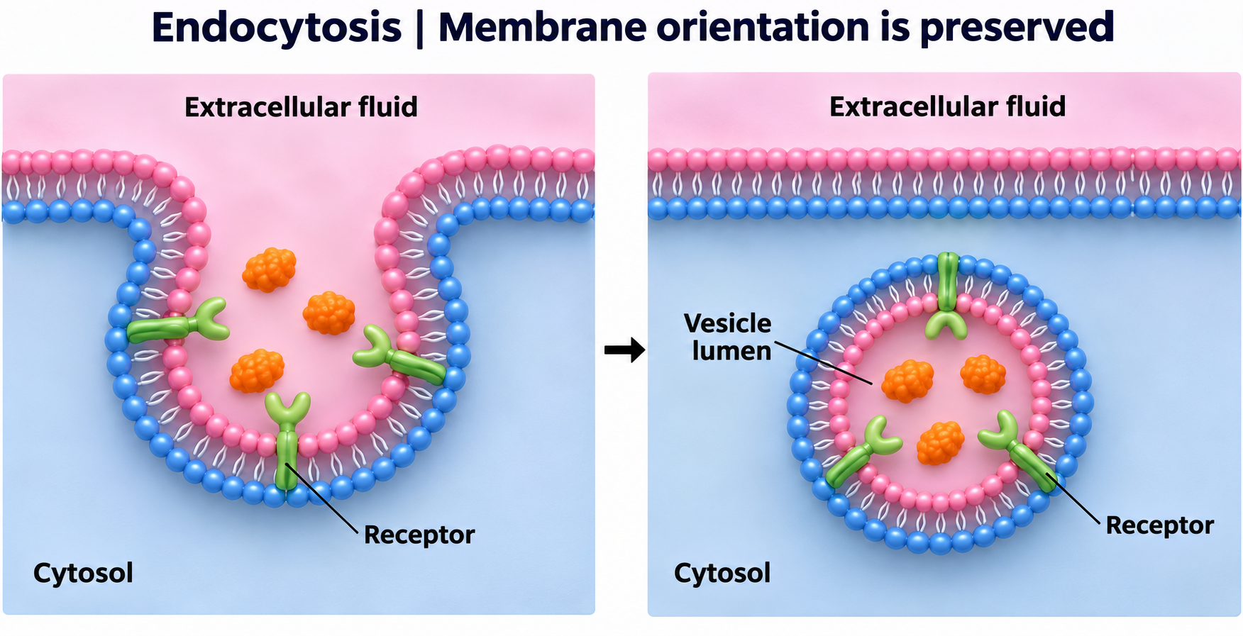
Membrane orientation
The extracellular-facing membrane surface becomes the lumen-facing surface of the vesicle.
Download original PNG
Budding and uncoating
A clathrin-coated pit becomes a coated vesicle and then sheds its coat.
Download original PNG