
Connect leaf tissues with light absorption, gas exchange and transport. The cross-sectional views give students a way to relate tissue position to the conditions each tissue experiences.
Choose from five images below. Open an image for a closer look, download its original PNG, or download all five as a ZIP.
Use these visuals in your lesson
Follow the path of carbon dioxide from outside the leaf to a photosynthetic cell. Then trace a separate path for water entering through the vascular tissue.
A question to discuss
Why are air spaces useful inside a leaf?
Leaf structure: five teaching visuals

The main layers
Upper epidermis, palisade mesophyll, spongy mesophyll and lower epidermis.
Download original PNG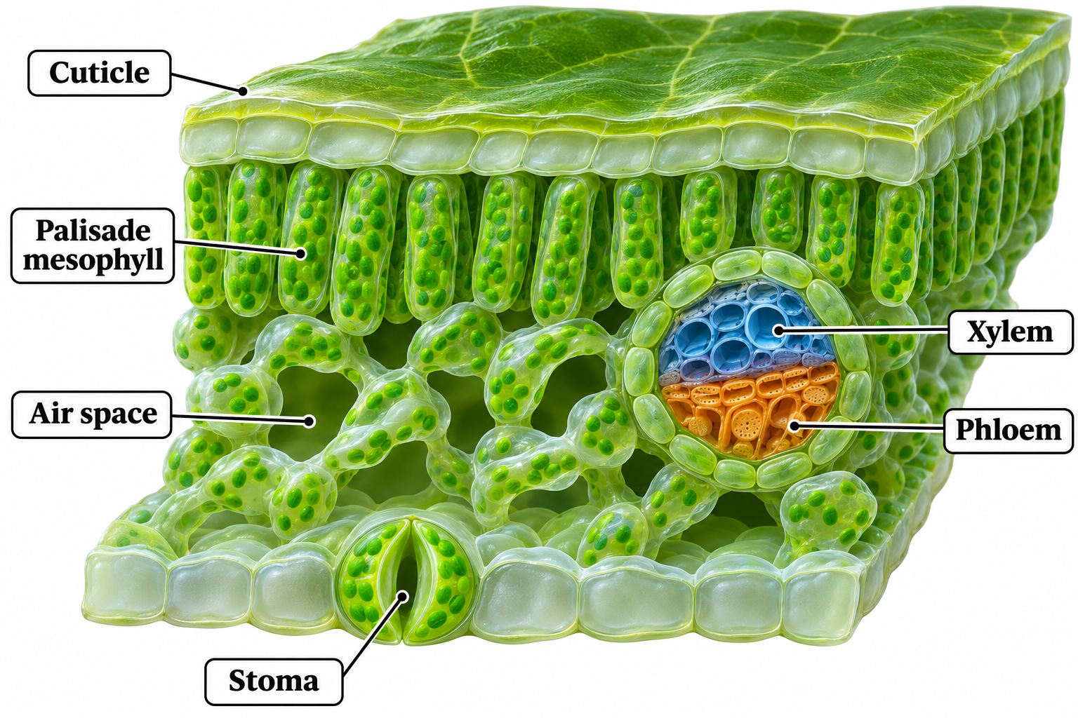
Inside the leaf
A cutaway showing cuticle, air spaces, stoma, xylem and phloem.
Download original PNG
A vascular bundle
Xylem, phloem and bundle sheath within a typical dicot leaf.
Download original PNG
Carbon dioxide enters
Follow a route from the stomatal pore through air spaces toward mesophyll cells.
Download original PNG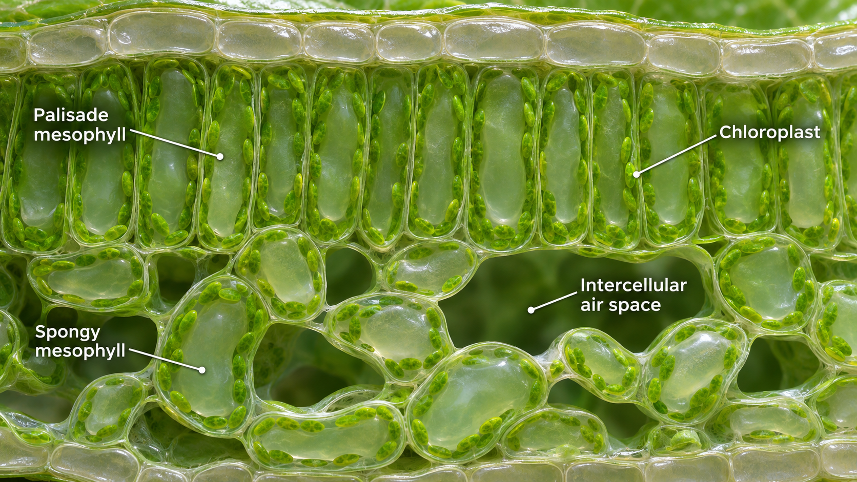
Mesophyll in detail
Palisade and spongy mesophyll, chloroplasts and intercellular air spaces.
Download original PNG