
Explain chromosome separation with a simplified chromosome model and selected views of the spindle and cytokinesis. The collection supports both the sequence of events and careful chromosome counting.
Choose from five images below. Open an image for a closer look, download its original PNG, or download all five as a ZIP.
Use these visuals in your lesson
Ask students what is being counted before giving a number. Compare chromosomes with DNA molecules so replication and chromatid separation are not confused.
A question to discuss
When sister chromatids separate, how does the chromosome count change within the still-undivided cell?
Mitosis: five teaching visuals
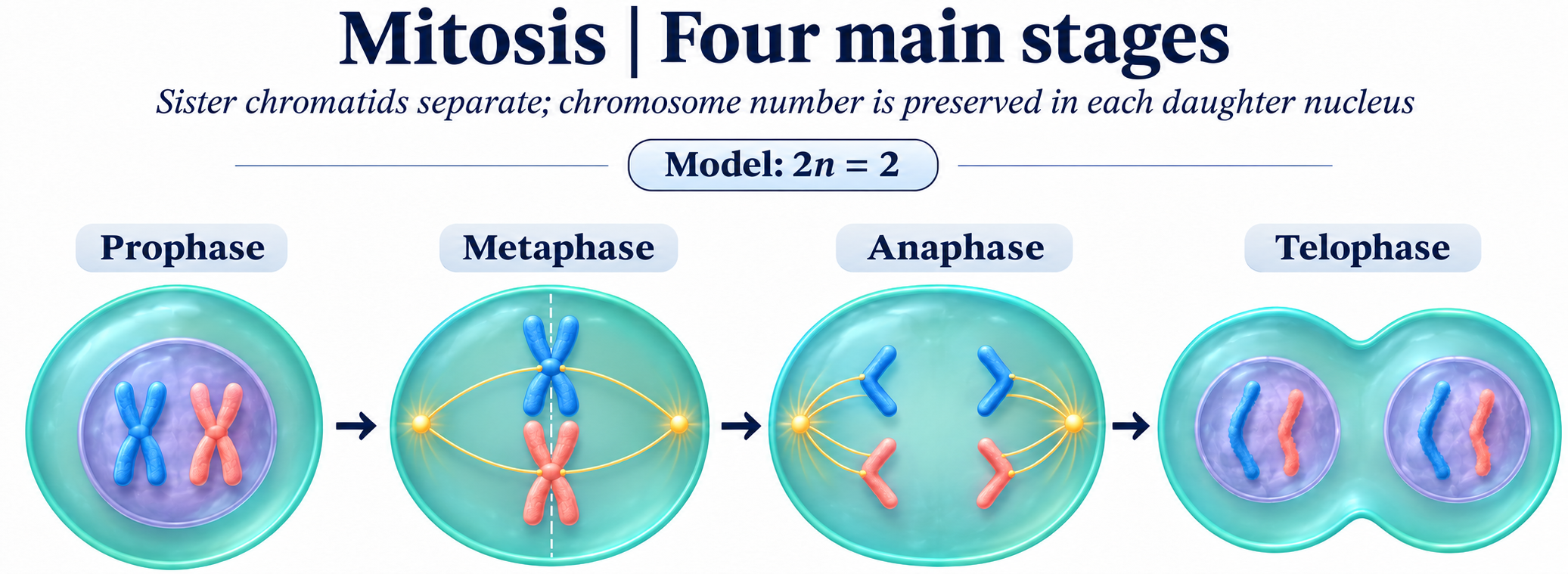
Four main stages
Prophase, metaphase, anaphase and telophase in a simplified 2n = 2 model.
Download original PNG
Metaphase attachment
Sister chromatids, kinetochores, spindle poles and the equatorial plane.
Download original PNG
One chromosome, two daughters
Track sister chromatids as they separate into daughter chromosomes.
Download original PNG
Cytokinesis
A cleavage furrow and contractile ring divide an animal cell after chromosome separation.
Download original PNG
DNA copied, then shared
Compare chromosome and DNA-molecule counts before replication and after division.
Download original PNG