
Explore the main regions of a neuron and the relationship between its structure and signalling role. Use the available views to introduce vocabulary before moving to electrical and synaptic mechanisms.
Choose from five images below. Open an image for a closer look, download its original PNG, or download all five as a ZIP.
Use these visuals in your lesson
Trace a signal through the represented neuron, naming regions as you go. Avoid treating one illustrated neuron as the only possible neuronal shape.
A question to discuss
How do the structures shown help a neuron receive and transmit information?
Neuron: five teaching visuals

The essentials
Dendrites, cell body, axon, myelin and terminals in a shortened neuron illustration.
Download original PNG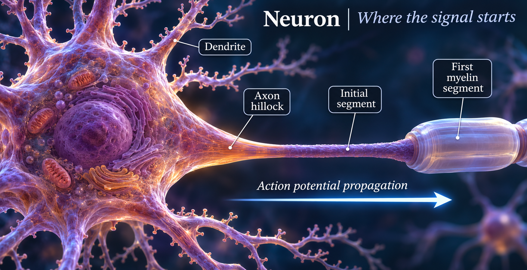
Where the signal starts
The axon hillock and initial segment lead into a myelinated axon.
Download original PNG
Insulation with gaps
Schwann-cell myelin surrounds a continuous axon, with nodes between segments.
Download original PNG
Node to node
Local current spreads beneath myelin and action potentials regenerate at nodes.
Download original PNG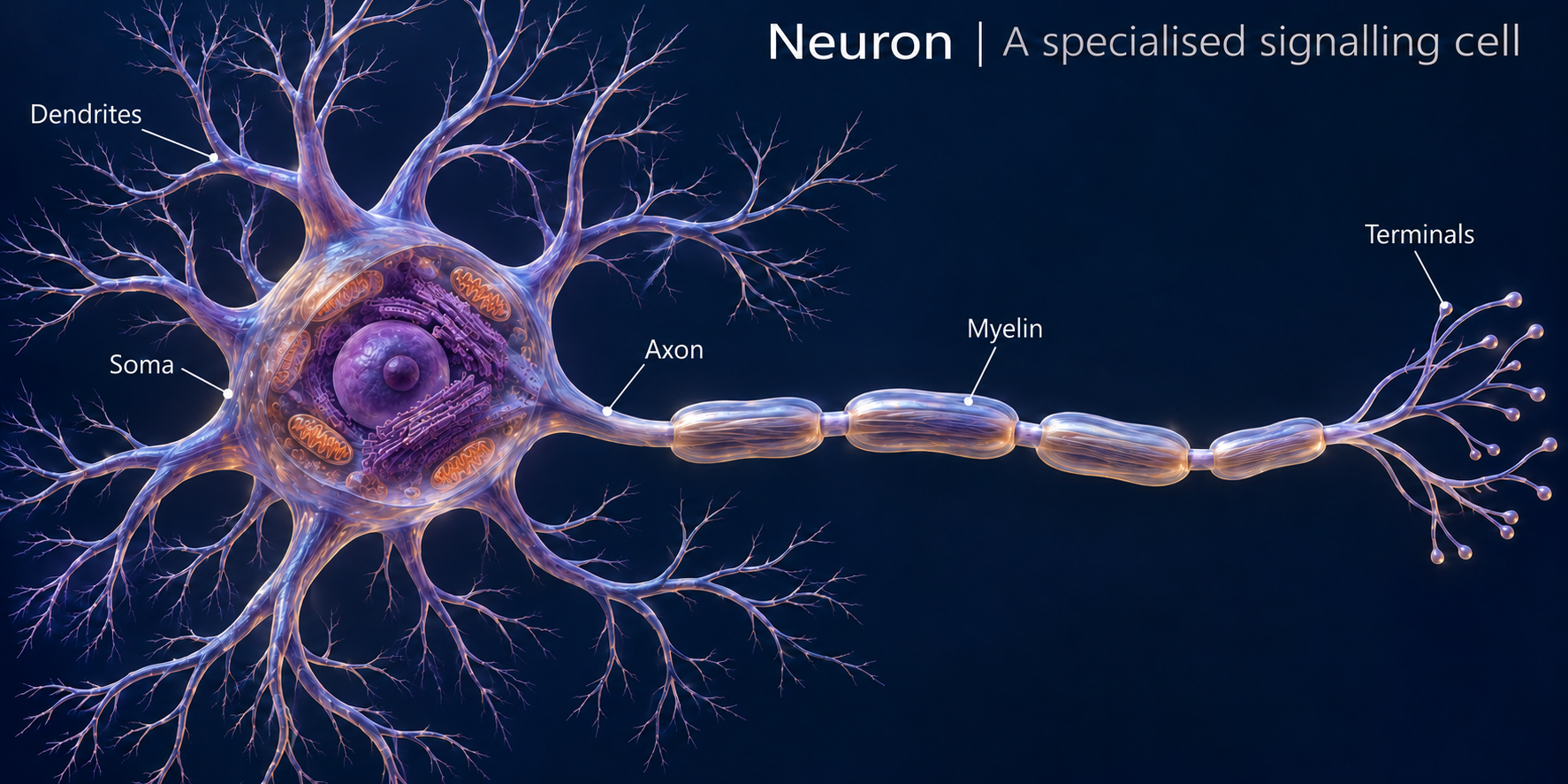
A specialised signalling cell
A detailed illustrative neuron with a shortened axon to show its features together.
Download original PNG