
Use heart visuals to connect chambers, valves and vessels with the movement of blood. Begin with the route through the heart before introducing pressure changes and the cardiac cycle.
Choose from five images below. Open an image for a closer look, download its original PNG, or download all five as a ZIP.
Use these visuals in your lesson
Trace one complete route through the pulmonary and systemic circuits. Ask students to justify the direction through each valve rather than memorising arrows alone.
A question to discuss
How do valves help maintain one-way flow through the heart?
The heart: five teaching visuals
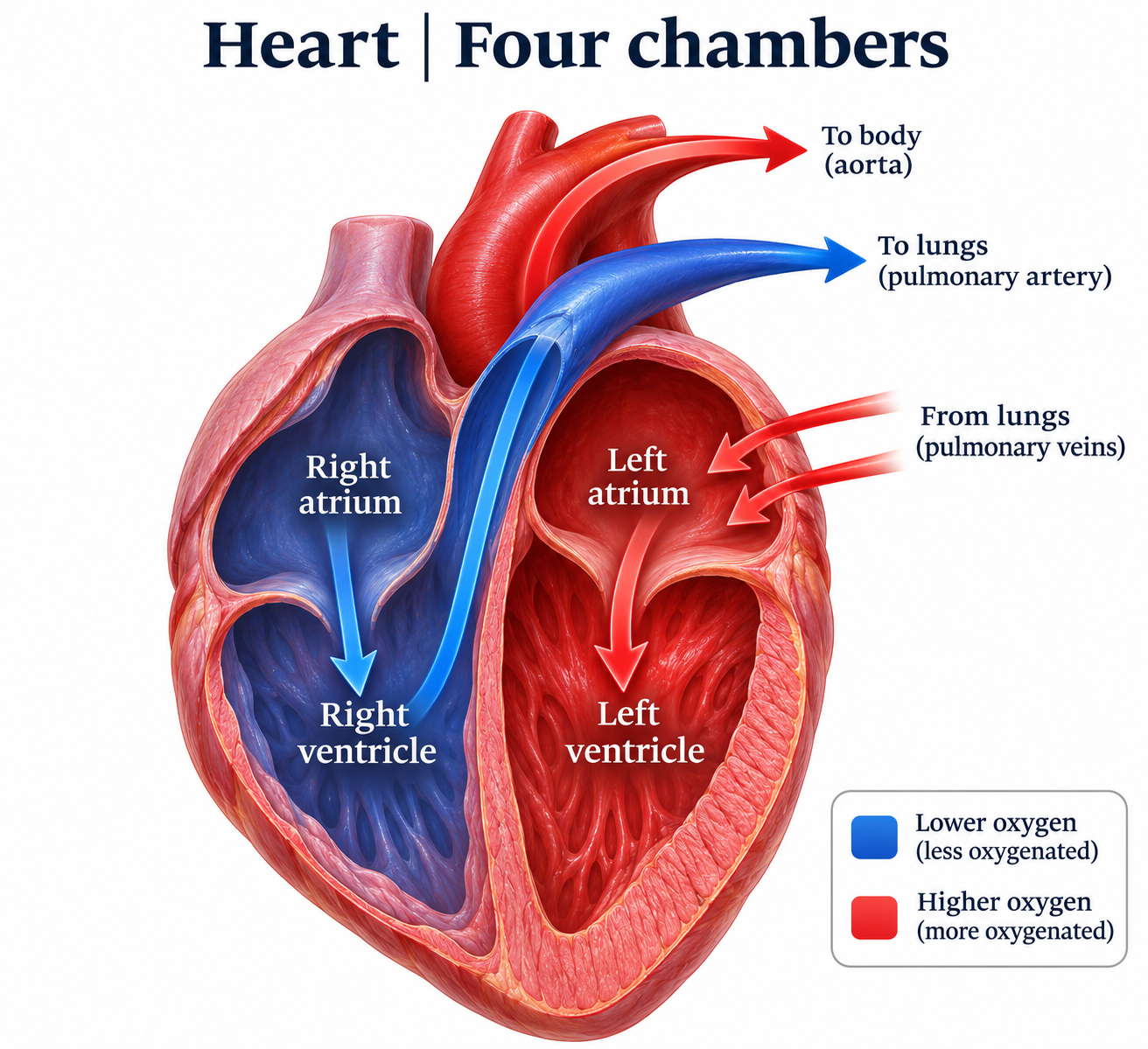
Four chambers
A heart cutaway follows blood through the right and left chambers to the lungs and body.
Download original PNG
Internal anatomy
A labelled cutaway identifies chambers, septum and major vessels.
Download original PNG
Valves guide the flow
Open-valve views compare pulmonary, aortic, tricuspid and mitral valve structures.
Download original PNG
Filling and ejection
Left-ventricular views compare valve positions during filling and ejection.
Download original PNG
One complete circuit
A numbered route follows blood through the heart, lungs and body tissues.
Download original PNG