
Follow fluid through a nephron while distinguishing filtration, reabsorption and secretion. The collection connects the tubule with its associated blood supply.
Choose from five images below. Open an image for a closer look, download its original PNG, or download all five as a ZIP.
Use these visuals in your lesson
Trace the filtrate and blood as two separate routes. Ask students to specify the direction of movement when they use the word reabsorption.
A question to discuss
Why is the composition of final urine different from the initial filtrate?
The nephron: five teaching visuals
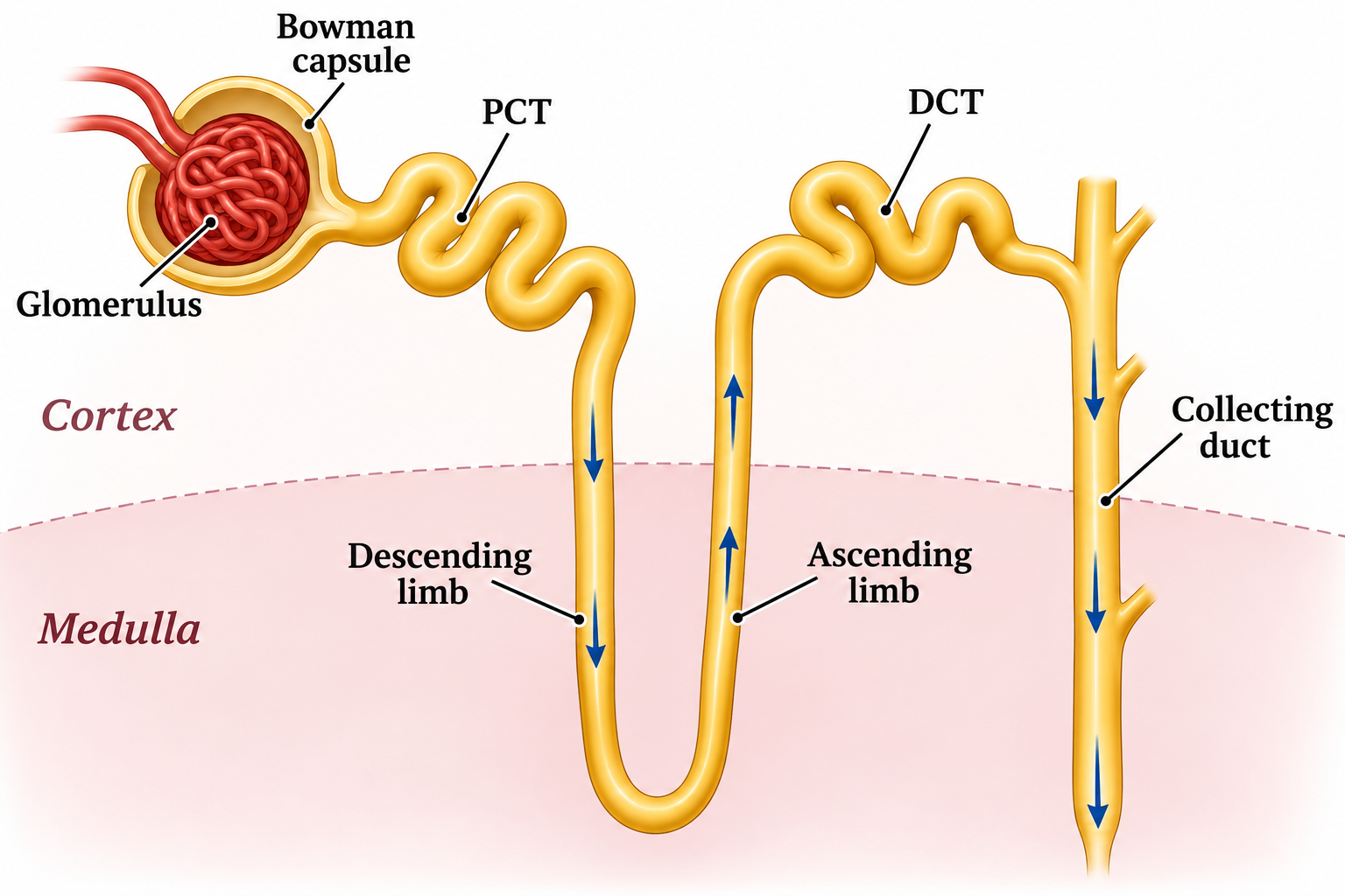
The filtrate route
A labelled nephron traces filtrate through the cortex and medulla into the collecting duct.
Download original PNG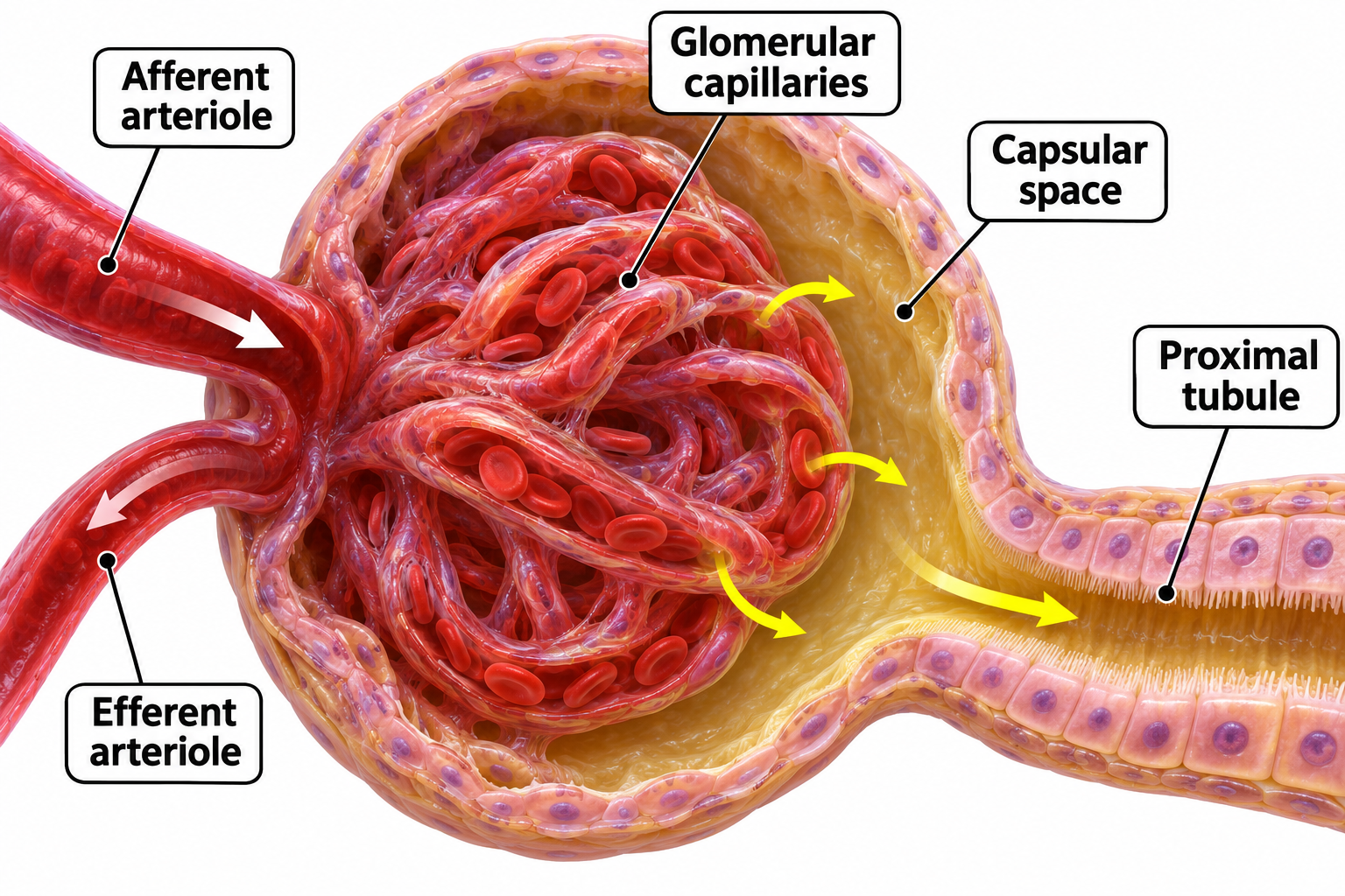
The renal corpuscle
A cutaway shows glomerular capillaries, arterioles, capsular space and the proximal tubule.
Download original PNG
The countercurrent loop
Descending and ascending limbs differ in water permeability and salt transport.
Download original PNG
Glucose reabsorption
Proximal-tubule transport links sodium–glucose cotransport, GLUT transport and the sodium–potassium pump.
Download original PNG
Filtration is only the start
A comparison follows water, glucose, urea and red blood cells in a healthy kidney.
Download original PNG