
Connect the intestinal surface with absorption into blood and lymph. These views move between larger folds, villi and the structures that increase contact with digested nutrients.
Choose from five images below. Open an image for a closer look, download its original PNG, or download all five as a ZIP.
Use these visuals in your lesson
Identify the scale of the view first so villi and microvilli are not confused. Trace a nutrient from the lumen across the epithelium and into an appropriate transport route.
A question to discuss
Why does a large epithelial surface help absorption?
Villi and absorption: five teaching visuals

More surface for absorption
Nested views distinguish intestinal folds, villi and the microvilli on epithelial cells.
Download original PNG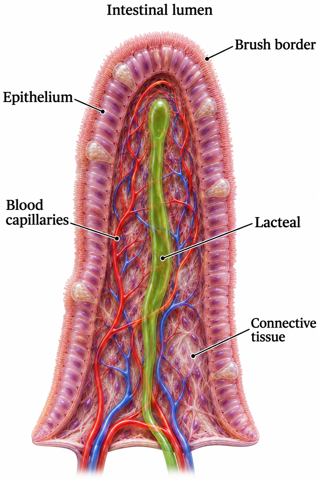
Inside a villus
A villus cutaway identifies epithelium, blood capillaries, lacteal and connective tissue.
Download original PNG
Microvilli
A brush-border close-up shows microvilli, actin bundles and the terminal web.
Download original PNG
Glucose into the blood
A simplified enterocyte model shows SGLT1, GLUT2 and the sodium–potassium pump supporting glucose absorption.
Download original PNG
Long-chain lipids into lymph
Absorbed lipid products are rebuilt and packaged into chylomicrons before entering a lacteal.
Download original PNG